Body Imaging

MBB’s dedicated team of body imaging radiologists specializes in the use of conventional and state-of-the-art techniques to diagnose and help treat diseases of the chest, abdomen, and pelvis. Our comprehensive services, provided in multiple Jacksonville-area hospitals and convenient outpatient settings, include x-ray, fluoroscopy, computed tomography (CT), ultrasound, magnetic resonance imaging (MRI), and positron emission tomography (PET-CT).

With advanced technologies allowing the evaluation of structures within the body like never before, MBB radiologists are able to diagnose disease and illness earlier and in a non-invasive manner.  This allows treatment to begin earlier and improves outcomes. Our body imaging radiologists work closely with your physicians to optimize your care—whether it be helping to select the best diagnostic test, accurately monitoring your course of treatment, or providing prompt, reliable results.

The 15 dedicated body imaging radiologists of MBB have vast depth and breadth of experience when it comes to evaluating diseases of the chest, abdomen, and pelvis. Our physicians have been educated and trained at many of the nation’s top medical centers, including the Cleveland Clinic, Duke, Harvard, Johns Hopkins, the Mayo Clinic, Washington University’s Mallinckrodt Institute of Radiology and the University of California, San Francisco.

When it comes to entrusting your care,  we invite you to take a closer look at the experience, commitment, and expertise that set apart the physicians of MBB. We also invite you to learn more about some of the different imaging studies performed by our experts in body imaging.

X-ray technology is the ‘workhorse’ of fundamental diagnostic imaging. Testing is fast, easy, and painless. Radiographs are produced by means of a source that radiates x-rays through a particular body part (i.e. spine). As the x-rays pass through the body they are weakened by the many types and levels of tissue encountered. That is why bony structures appear more prominently (i.e. whiter) than soft tissue – bone is denser and absorbs a greater amount of radiation. The energy is directed into a film cassette that has been placed into a receptacle under/behind the targeted body part. The film is exposed and developed in much the same manner as a photograph.

Computed Tomography (CT)

Computed tomography (CT) uses special x-ray equipment to create cross-sectional images of the body. Sometimes called a CAT scan, this technique is able to depict the internal organs with exquisite detail. Normal and pathologic tissues can be identified throughout all organ systems, including the brain, lungs, bones, abdominal organs, and blood vessels with great clarity.

What is CT? How Does it Work?

During a CT scan, multiple x-ray beams and highly sophisticated computer processors are used to create a series of images that show the inside of the body in cross section— much like slices in a loaf of bread. These images are able to provide much greater anatomic detail than conventional x-rays. As the technology behind CT scanning has improved since the first scanner was introduced in 1982, the multi-slice scanners now utilized at all MBB facilities are able to image the entire body in less than 18 seconds. The speed of these exams allows this technology to be especially useful in the setting of trauma, where the rapid detection of life-threatening injuries saves lives on a daily basis.

What is CT Used For?

As a diagnostic tool, the uses of CT are many and constantly increasing. CT is a mainstay of oncologic (cancer) imaging—both in cancer detection and in evaluating the success of various therapies. CT provides a highly accurate, non-invasive means of assessing the blood vessels, including the aorta in patients who may have an aneurysm or the renal arteries in patients who may have a narrowing contributing to an elevation in blood pressure.

CT is the most sensitive means of evaluating the urinary system for stones, and a newer technique, CT urography, has become the standard of care in the initial evaluation for potential tumors of the urinary tract. Infectious processes and other inflammatory conditions are well-suited to CT evaluation, and the technique is often used to guide needle biopsy, radiation therapy, fluid drainage, and other minimally invasive procedures.

What to Expect During a CT Exam

CT scans are painless, but they do require you to lay still for anywhere between 10 seconds and a few minutes. For the exam, you will lie on a padded scanning bed. While the scan is performed quickly, you may spend between 5 and 30 minutes on the scanning bed, depending on the type and number of exams you are having. While most patients lie on their backs, your position may vary depending on the type of CT exam you are having. The technologist performing the study will not stay in the room during the scan, but you can speak with him or her by intercom throughout the exam.

During the scan, you will hear normal whirring and mechanical noises as the scanner rotates around your body. You may be asked to hold your breath for short periods of time. It is important not to move any part of your body during the scan in order to avoid blurring the images. This is similar to having your photo taken.

In order to provide greater diagnostic information, CT scans are often performed with intravenous contrast. This requires placement of an IV by the technologist, followed by injection of an iodine-based “dye” shortly before the images are obtained. Please note that the contrast may make you feel warm or create a metallic taste in your mouth for a short time. Some CT scans are best performed after drinking a flavored oral contrast to provide better visualization of the gastrointestinal tract.

How to Prepare for a CT Exam

Wear comfortable, loose clothing that does not have zippers or other metal fasteners. Depending upon the type of CT scan, you may be asked to change into a hospital gown prior to the exam.

If your CT scan requires that you receive IV contrast, please refrain from eating or drinking 4 hours prior to the exam. (You may take prescribed medications with small amounts of liquid.) Depending upon your age and health, you may be asked to undergo a quick blood test to check your kidney function. If your exam requires oral contrast, this will be provided at the facility where you check in, or it may be provided at your physician’s office.

Before the exam, a CT technologist will ask you a few medical questions, explain your procedure, and assist you into the CT examination room.

For all CT exams, it is important to let the technologist know of any of the following:

  • If you are pregnant or think there is a chance you could be pregnant
  • If you are allergic to latex, iodine, or any medication
  • If you have had a previous allergic reaction to contrast
  • If you have asthma
  • If you have kidney disease or renal failure
  • If you are diabetic and what medications you take
  • If you ate or drank before the test

You may return to your normal activities immediately after the scan.

The CT images obtained during your exam will be reviewed and interpreted by an MBB radiologist, and a report of his or her findings will be provided to your physician.

MRI

Magnetic Resonance Imaging (MRI)

Magnetic resonance imaging (MRI) is a non-invasive technique utilizing a strong magnet together with radio waves and complex computer processing to produce unparalleled high resolution images of the internal organs and soft tissues of the body. Unlike traditional radiologic techniques, MRI does not use radiation or x-rays to obtain images. The exceptionally clear and detailed images allow the physicians of MBB to detect subtle differences between normal and diseased or injured tissue.

What is MRI? How Does it Work?

During an MRI scan, the patient is exposed to a strong magnetic field and painless radiofrequency waves. These waves enter the body and then echo back to a special receiver coil placed on the part of the body being imaged. The returning waves contain information about the inside of the body, and sophisticated computer processing turns that information into images with exquisite anatomic detail.

What is MRI Used For?

While MRI has revolutionized musculoskeletal and neurologic imaging, it has also become a valuable adjunct to computed tomography (CT) in imaging of the chest, abdomen, and pelvis.

Within the chest, some of the latest advances in MRI are being used to image the heart. MRI is very effective in evaluating how much cardiac tissue has been damaged in a heart attack, and this information can help determine if patients should undergo bypass surgery. The information from this exam can also be used to create a “movie” showing the beating heart in motion. This allows the radiologist to evaluate whether the actual function of the heart is impaired and to what degree. MRI is also frequently used to evaluate the aorta and other blood vessels.

In the abdomen, MRI shines in its ability to definitively characterize lesions of the liver, pancreas, kidneys, and adrenal glands. In many cases, MRI can determine if tumors are benign or malignant without the need for surgery or a biopsy. If a tumor is found to be malignant, the radiologist can use the images to help the surgeon plan the safest possible operation.

Within the pelvis, MRI is very effective in evaluating the uterus in patients suspected to have fibroids or other conditions causing pain or abnormal bleeding. MRI of the pelvis also provides valuable diagnostic information in the setting of infertility, pelvic floor dysfunction, or cancers of the ovaries, cervix, prostate, or rectum. In many cases, the radiologist uses detailed MRI images to assist treating surgeons and oncologists in deciding the best course of treatment for pelvic malignancies.

MR angiography (MRA) is a sensitive, non-invasive means of evaluating the blood vessels. Though most commonly used to study the arteries and veins supplying and draining the brain, this technique may be applied to almost any vessel in the body. While MRA traditionally required the injection of a “dye” into the veins, newer techniques have been shown to be almost just as accurate without the need for a contrast injection.

What to Expect During an MRI Exam

Before your exam, a specially trained MRI technologist will discuss the exam with you, review your medical history, and answer any questions you may have. He or she will be in contact with you throughout the exam, keeping you informed and providing support.

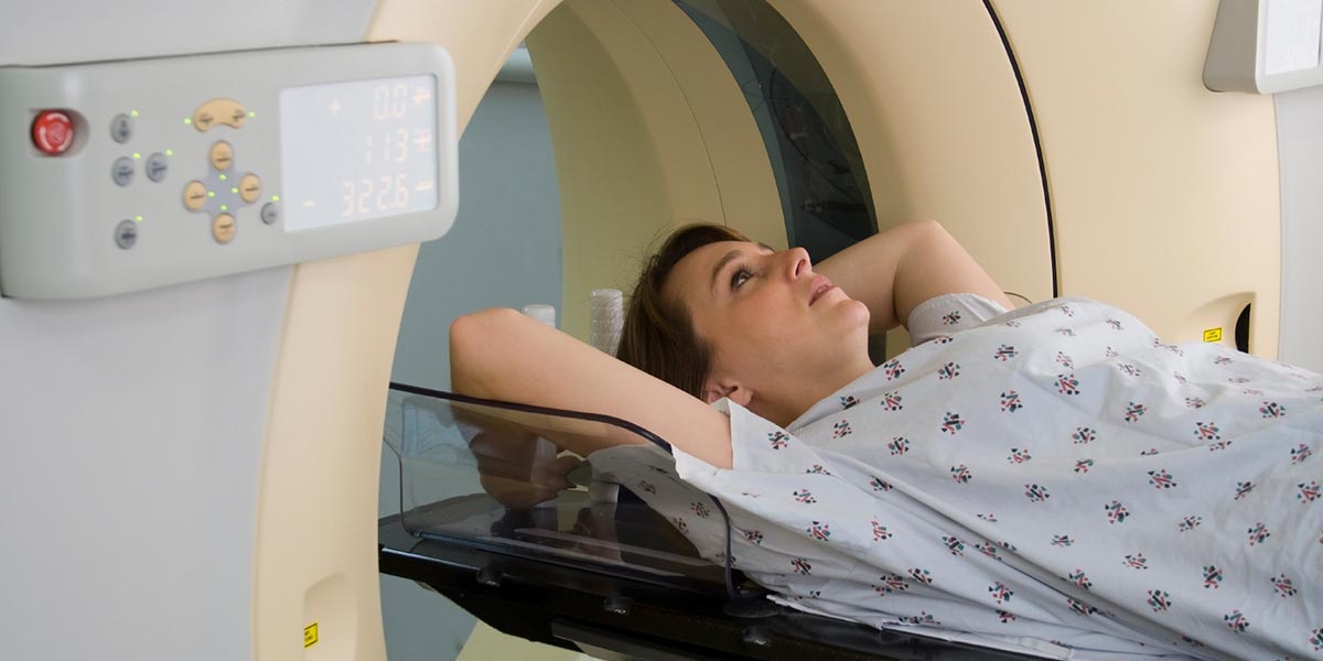
MRI exams are painless, but they do require you to lay fairly still for 20-45 minutes. For the exam, you will lie on a padded scanning bed. While most patients lie on their backs, your position may vary depending upon the type of exam and body part being imaged. The technologist performing the study will not stay in the room during the exam. He or she will be in a control room operating the MRI scanner. The technologist will be watching you through a glass window, and you will be able to communicate by intercom throughout the exam.

When images are being acquired, the MRI scanner makes loud “knocking” noises—caused by the rapidly shifting magnetic fields. You will be given earplugs to dampen the noise, and you may be given headphones to listen to the music of your choosing.

You may be asked to hold your breath for short periods of time, and you will be asked to hold as still as possible for intervals of several minutes at a time. During these periods, the scanner is creating pictures of your internal organs. Much like a photograph, movement will cause the image to blur—making it more difficult for your doctors to diagnose and treat your condition.

In order to provide greater diagnostic information, some MRI scans are performed with intravenous contrast. This dye is made with the element gadolinium—a relatively inert compound with a very low incidence of allergic reaction. For certain exams of the bowel and bile ducts, you may be asked to drink an oral contrast agent shortly before the exam.

How to Prepare for an MRI Exam

Wear comfortable, loose clothing that does not have zippers or other metal fasteners. If this is not possible, you may be asked to change into a gown so that metal on your clothing does not interfere with the MRI. If this is the case, you will be shown to a private changing room.

Before the exam, a technologist will ask you a few medical questions and explain your exam before assisting you into the MRI scanning room.

MRI is very safe, and there are no health risks associated with the magnetic field or the radio waves used by the machine. However, there are some special circumstances that limit the use of strong magnetic fields, so it is important that you let the technologist know if you have any of the following:

  • Cardiac pacemaker, internal defibrillator, stent, or artificial heart valve.
  • Insulin or other infusion pump.
  • Aneurysm clip.
  • Cochlear implant
  • Artificial joint, metallic plate, pin, or other metallic implant.
  • Intrauterine device

While some of the above medical devices do not present a problem, others are not compatible with the MRI scanner. If you have any of the above devices, you may want to call the department before your appointment to ensure that you are a candidate for an MRI exam. If possible, please bring any information you may have about your implantable medical device.

For all MRI exams, it is also important to let the technologist know of any of the following:

  • If you are pregnant or think there is a chance you could be pregnant.
  • If you are allergic to any medications or intravenous contrast.
  • If you have kidney disease or renal failure.
  • If you have liver disease.
  • If you have a history of metalworking or have ever had a fragment of metal get in your eye.
  • If you have a history of prior gunshot wound.
  • If you have permanent (tattoo) eye-liner.

Please note that pregnant women may undergo MRI examinations; however, they should discuss the exam with their ordering physician and/or radiologist before the study. While there are no known adverse effects of MRI scanning on the developing baby, it is recommended that imaging not be performed until the second trimester, if possible.

While the images are being obtained, you will be lying in a tube that creates the magnetic field. Over the years, advances in technology have allowed these tubes to be made wider; however patients with claustrophobia may still feel uncomfortable and unable to tolerate the exam. If you are prone to claustrophobia or panic attacks, please discuss this with your physician. He or she may wish to prescribe an anti-anxiety medication for you to take prior to your exam. In our experience, most claustrophobic patients are then able to complete an MRI without difficulty. If you do take such a medication before your exam, please be sure a friend or family member comes with you, as you will need someone to drive you home.

After complex computer processing, data from your MRI exam will be used to create hundreds, or even thousands, of detailed images. These images will be analyzed and interpreted by an MBB radiologist, and a report of his or her findings will be provided to your physician.

Ultrasound (US) imaging utilizes high frequency sound waves to evaluate structures within the body. A small hand-held probe is pressed against the skin, emitting sound waves that cannot be heard. These waves are reflected by different tissues within the body, and advanced computer processing is used to create images of the inside of the body. Unlike conventional x-rays and CT scanning, sonography does not use 
radiation, making it a favorite imaging modality in pregnant women and in the pediatric population.

Ultrasound is frequently used to image the abdominal organs, including the liver, pancreas, spleen, and gallbladder when patients have pain or laboratory abnormalities. Ultrasound remains the most sensitive test for finding gallstones, and it is a fast, non-invasive way of evaluating the kidneys and bile ducts for obstruction. It is excellent for differentially cystic from solid structure in the body.

While US visualization of structures deep within the body may be limited by excessive body fat or gas within the bowel, ultrasound is highly effective at evaluating superficial structures, including the thyroid gland, the breasts, and the testicles.

An advantage of ultrasound is its ability to detect and analyze motion. This allows for evaluation of blood flow within arteries, veins, and surgically-created grafts. Ultrasound is commonly used to assess the veins of the legs to look for potentially lethal blood clots which could dislodge and migrate to the lungs. Ultrasound is also frequently used to screen for dangerous enlargement of the aorta (aneurysm) in at-risk populations.

Ultrasound is the initial imaging modality of choice for evaluation of the female pelvis—providing a detailed look at the uterus and the ovaries. Perhaps the best known application of ultrasound is in the evaluation of the pregnant woman. US is well-suited to evaluate the growing fetus, surrounding amniotic fluid, and the placenta. Ultrasound may be utilized to evaluate fetal size, position, movement and heart rate. In addition to imaging with a probe pressed against the skin of the lower abdomen, some examinations of the female pelvis require use of a transvaginal probe, which the patient inserts into the vaginal canal. This allows for much more detailed imaging of the uterus and ovaries, particularly in patients who are not pregnant or are in the early stages of pregnancy.

What is nuclear medicine?

Nuclear medicine uses small amounts of short-lived radioactive material to diagnose or treat diseases and to give information on how tissues and organs work. Nuclear medicine can detect diseases such as heart disease, arthritis, and cancer. Areas of the body most often studied include the brain, thyroid gland, heart, lungs, kidneys, gallbladder, liver, and bones.

How does nuclear medicine work?

Before having a nuclear medicine procedure performed, patients are given a radioactive material called an isotope. Isotopes can be injected, inhaled, or swallowed in liquid or capsule form. Once the isotope is inside the body, it travels to the target organs or tissues and gives off gamma rays. Images are then taken of the body with special equipment that can detect the gamma rays. These images are interpreted by a radiologist with special training in nuclear medicine.

What kind of equipment is used for nuclear medicine studies?

Various types of equipment are used for nuclear medicine studies depending on the procedure. Gamma probes, stationary gamma cameras, and Single photon emission computed tomography (S PECT) cameras. Nuclear medicine probes look like long metal tubes. They can take images of the body by being waved over the patient. Nuclear Medicine probes are not inserted into the body and usually do not come in direct contact with the skin. Stationary gamma cameras are attached to a tall unit and positioned very close to the area being studied. S PECT cameras can rotate around the patient to take the images.