Musculoskeletal Imaging

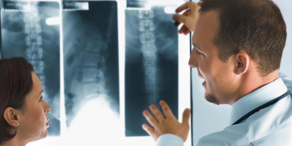
MBB Radiology provides complete musculoskeletal (MSK) imaging and intervention services. Our service includes both conventional and state-of-the-art techniques such as imaging guided interventional procedures. Our MSK imaging team has decades of combined experience.Each of our subspecialty MSK radiologists is fellowship trained, providing a full complement of physicians with extensive experience and special interest in musculoskeletal and body imaging.

Included are physicians who have targeted training in sports medicine, body imaging, complicated arthritis and rheumatology, as well as orthopedic tumors. Our MSK radiologists have been educated and trained at many of the country’s most respected institutions including the Mayo Clinic, Harvard, Duke, Vanderbilt, and Washington University. When it comes to your care, take a closer look at who is providing the interpretation of your imaging study and have confidence in MBB Radiology.

In addition to treating you and your family we also provide expert image interpretation for the Jacksonville Jaguars, a National Football League team, as well as many other professional and amateur athletes in Northeast Florida.

Musculoskeletal imaging is not a one-size-fits-all specialty. The choice of one or more imaging examinations depends on the specific needs of our patient. MBB radiologists are routinely in consultation with your doctors to discuss your case and recommend the most appropriate test. At MBB radiology you can rest assured in our experience and commitment to your care.

Conventional Radiography or X-ray is an imaging technique that has been used since 1895 to show up abnormalities in bones and certain body tissue, such as breast tissue.

X-rays are a type of high-energy radiation that is like light waves but higher in energy. An X-ray machine can produce short bursts of X-rays that pass easily through fluids and soft tissues of the body but are blocked by dense tissue such as bone.

Contrast X-rays use a substance (called a contrast medium) that makes hollow or fluid-filled structures visible. This means that structures such as the digestive tract, blood vessels or urinary system that do not usually show up on X-ray, can be seen. The substance is injected or swallowed and X-rays cannot pass through it, so the area will appear white on the X-ray.

Fluoroscopy is a type of medical imaging that shows a continuous x-ray image on a monitor, much like an x-ray movie. It is used to diagnose or treat patients by displaying the movement of a body part or of an instrument or dye (contrast agent) through the body. During a fluoroscopy procedure, an x-ray beam is passed through the body. The image is transmitted to a monitor so that the body part and its motion can be seen in detail.

Uses

  • Fluoroscopy is used in many types of examinations and procedures. Some examples include
  • Barium x-rays and enemas (to view movement through the GI tract)
  • Catheter insertion (to direct the placement of a catheter during angioplasty or angiography)
  • Blood flow studies (to visualize blood flow to organs)
  • Orthopedic surgery (to view fractures and fracture treatments)

Risks/Benefits

Fluoroscopy is a type of x-ray procedure, and it carries the same types of risks as other x-ray procedures. The radiation dose the patient receives varies depending on the individual procedure.

  • The two major risks associated with fluoroscopy are:
  • Radiation-induced injuries to the skin and underlying tissues (“burns”)
  • The small possibility of developing a radiation-induced cancer some time later in life.

When an individual has a medical need, the benefit of fluoroscopy far exceeds the small cancer risk associated with the procedure. Even when fluoroscopy is medically necessary, it should use the lowest possible exposure for the shortest possible time.

MRI


Unlike traditional radiologic techniques, MRI (Magnetic Resonance Imaging) does not use radiation or x-rays to obtain images. MRI utilizes a strong magnetic field, radio waves, and complex computer processing to produce unparalleled high resolution images of the internal organs and soft tissues of the body. This painless examination takes approximately 30 to 60 minutes to complete depending on the part of the body being imaged. While MRI has revolutionized musculoskeletal and neurologic imaging, it has also become a valuable adjunct to computed tomography (CT) in imaging of the chest, abdomen, and pelvis. Within the chest, MRI is frequently utilized to evaluate the aorta and the musculature of the heart, as well as to assess the complex bundle of nerves in the axillary region known as the brachial plexus. In the abdomen, MRI shines in its ability to definitively characterize lesions of the liver, pancreas, kidneys, and adrenal glands. MR cholangiopancreatography (MRCP) allows for non-invasive evaluation of the bile ducts draining the liver. A common application of pelvic MRI is for evaluation of the uterus in patients suspected to have fibroids or other conditions causing pain or abnormal bleeding. MRI of the pelvis may also be helpful in the setting of infertility, pelvic floor dysfunction, or cancers of the ovaries, cervix or prostate. MR angiography (MRA) is a sensitive, non-invasive means of evaluating the blood vessels. Though most commonly used to study the arteries and veins supplying and draining the brain, this technique may be applied to almost any vessel in the body.

CT

Computed tomography (CT) uses special x-ray equipment to create cross-sectional image of the body. Sometimes called a CAT scan, this technique is able to depict the internal organs with exquisite detail. Normal and pathologic tissues can be identified throughout all organ systems including lung, bone, brain, soft tissue, and blood vessels with great clarity. CT scans are painless and noninvasive. Most exams can be completed within a few minutes. The speed of these exams allows this technology to be especially useful in the setting of trauma, where life-threatening injuries may be detected within minutes.

Joint injections or aspirations (taking fluid out of a joint) usually are performed with a cold spray or other local anesthesia in the office or hospital setting. After the skin surface is thoroughly cleaned, the joint is entered with a needle attached to a syringe. At this point, either joint fluid can be obtained (aspirated) and used for appropriate laboratory testing or medications can be injected into the joint space. This technique also applies to injections into a bursa or tendon sheath to treat bursitis and tendonitis, respectively.

Joint injections may decrease the accumulation of fluid and cells in the joint and may temporarily decrease pain and stiffness. They may be given to treat inflammatory joint conditions, such as rheumatoid arthritis, psoriatic arthritis, gout, tendonitis, bursitis and, occasionally, osteoarthritis.

Corticosteroids (such as methylprednisolone and triamcinolone formulated to stay primarily in the joint) frequently are used. They are anti-inflammatory agents that slow down the accumulation of cells responsible for producing inflammation and pain within the joint space. Although corticosteroids may also be successfully used in osteoarthritis, their mode of action is less clear. Hyaluronic acid is a viscous lubricating substance that may relieve the symptoms of osteoarthritis of the knee for periods up to 6–12 months. Mode of action is not clear.

Joint aspiration usually is done for help with diagnosis or treatment. Fluid obtained from a joint aspiration can be examined by the physician or sent for laboratory analysis, which may include a cell count (the number of white or red blood cells), crystal analysis (to confirm the presence of gout or pseudogout), and/or culture (to determine if an infection is present inside the joint). Drainage of a large joint effusion can provide pain relief and improved mobility. Injection of a drug into the joint may yield complete or short-term relief of symptoms.


Arthrography is medical imaging to evaluate conditions of joints. In addition to its diagnostic role, we uses fluoroscopic guidance to inject steroids and anesthetic into joint space, resulting in significant pain relief and an increase in joint function for selected patients.

Conventional arthrography is the x-ray examination of a joint that uses a special form of x-ray called fluoroscopy and an injection of contrast material containing iodine directly into the joint. Alternate methods of arthrography examinations use magnetic resonance imaging (MRI) or computed tomography (CT).

An x-ray (radiograph) is a noninvasive medical test that helps physicians diagnose and treat medical conditions. Imaging with x-rays involves exposing a part of the body to a small dose of ionizing radiation to produce pictures of the inside of the body. X-rays are the oldest and most frequently used form of medical imaging.

Fluoroscopy makes it possible to see bones, joints and internal organs in motion. When iodine contrast is injected into the joint, it fills the entire joint and becomes clearly visible during x-ray evaluation, allowing the radiologist to assess the anatomy and function of the joint. Although the injection is typically monitored by fluoroscopy, the examination also involves taking radiographs for documentation. The images are most often stored and viewed electronically.

Similarly, MR arthrography also involves the injection of a contrast material into the joint. The contrast material used for MR evaluation is different from that used for x-ray; it contains gadolinium, which affects the local magnetic field within the joint. As in conventional arthrography, the contrast material outlines the structures within the joint and allows them to be evaluated by the radiologist after the MR images are produced.

MRI uses a powerful magnetic field, radiofrequency pulses and a computer to produce detailed pictures of organs, soft tissues, bone and virtually all other internal body structures. The images can then be examined on a computer monitor, printed or copied to CD. MRI does not use ionizing radiation (x-rays).

CT arthrography uses the same type of contrast material as conventional arthrography and may be supplemented by air to produce a double contrast CT arthrogram. CT makes cross sectional images processed by a computer using x-rays. Once the joint space is localized with one of these methods, anesthetic or steroids can be administered.

Vertebroplasty and kyphoplasty are minimally-invasive procedures that are used to treat vertebral fractures, usually caused by osteoporosis or bone tumors. The pain of vertebral fractures is caused by the compression and shifting of the damaged vertebrae, so vertebroplasty and kyphoplasty treat the pain and the fracture by shoring up the vertebrae with bone cement.

Vertebroplasty and kyphoplasty are minimally invasive procedures in which fluoroscopy (live x-ray) imaging is used to precisely place a needle at the location of the fracture. The bone cement is injected directly into the vertebrae, and then allowed to harden so it can strengthen and support the vertebrae.

Vertebroplasty is appropriate for cracked or broken vertebrae, but for vertebrae that have suffered compression fractures, kyphoplasty is more successful. Kyphoplasty involves the insertion of a balloon catheter into the compressed vertebrae. The balloon is filled with bone cement and allowed to harden. This restructures and rebuilds the vertebrae so that it can provide support for the spine again.

Both vertebroplasty and kyphoplasty not only relieve the pain of damaged vertebrae, but they also prevent further damage from the original fracture.

We also offer RF Kyphoplasty. This procedure utilizies ultra-high viscosity cement and a directional device which allows for precise placement of the cement into tumors and fractures.

It requires no stitches, no general anesthesia, and it has a very quick recovery time. Most people experience complete pain relief immediately after the procedure, others report that their pain was gone or significantly reduced within 48 hours.

This is a needle-based procedure that our doctors perform at our hospital and outpatient facilities. The procedure takes about an hour and requires no overnight stay.